Unlock Knee Pain: Understanding Essential Meniscus Tear Tests
Experiencing persistent knee pain, swelling, or a frustrating popping sensation? These could be tell-tale signs of a meniscus tear, a common yet debilitating injury that affects over a million Americans each year. Understanding the diagnostic process, particularly the various physical examination techniques, is crucial for an accurate diagnosis and effective treatment. This comprehensive guide will delve into the world of the meniscus tear test, exploring how healthcare professionals identify this often-elusive injury.
Navigating the complexities of knee pain can be daunting, but pinpointing the exact cause is the first step towards recovery. While advanced imaging like MRI plays a significant role, the initial assessment often relies on a series of clinical tests designed to evaluate the integrity of your knee's vital structures. These tests, performed by skilled practitioners, are invaluable tools in the diagnostic arsenal, helping to differentiate a meniscus tear from other knee ailments.
Table of Contents
- The Meniscus: Your Knee's Crucial Cushion
- Understanding Meniscus Tears: A Common Injury
- Why Accurate Diagnosis Matters: The Role of Meniscus Tear Tests
- The Cornerstone: McMurray's Test Explained
- Beyond McMurray: Other Key Meniscus Tear Tests
- The Diagnostic Journey: Clinical Tests to Advanced Imaging
- What to Expect After a Meniscus Tear Diagnosis
- Empowering Your Recovery: Seeking Expert Care for Meniscus Tears
The Meniscus: Your Knee's Crucial Cushion
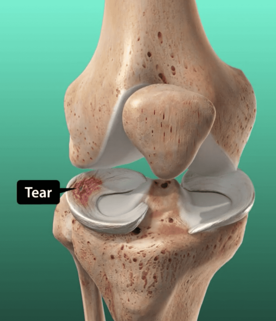
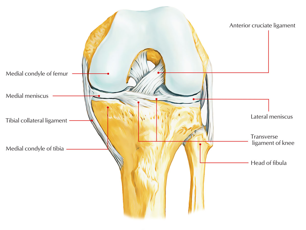
Before diving into the diagnostic tests, it's essential to understand what the meniscus is and why it's so vital for knee health. Your knee joint, a marvel of biomechanical engineering, relies on several components to function smoothly and absorb impact. Among the most critical are the menisci (plural of meniscus). You have two C-shaped pieces of cartilage in each knee: the medial meniscus (on the inner side of the knee) and the lateral meniscus (on the outer side).
- Jayshree Gaikwad Web Series
- Xxx Is Equal To 2025
- Jayshree Gaikwad Movies
- The Secret Life Of A Campus Wife Manhwa
- Wwxx Lyrics
These cartilaginous pads act like shock absorbers, distributing weight evenly across the knee joint and preventing direct bone-on-bone contact between the femur (thigh bone) and the tibia (shin bone). They also contribute to knee stability and lubrication, facilitating smooth movement. Without healthy menisci, the knee joint is vulnerable to increased stress, leading to pain, instability, and accelerated wear and tear of the articular cartilage, potentially resulting in osteoarthritis. Understanding their role underscores why accurately diagnosing a torn meniscus is so important.
Understanding Meniscus Tears: A Common Injury
Meniscus tears are incredibly common, affecting people of all ages and activity levels. As mentioned, over 1 million Americans tear their meniscus each year, highlighting the prevalence of this injury. Tears can occur in various ways. In younger, more active individuals, they often result from acute trauma, such as twisting the knee while the foot is planted, common in sports like soccer, basketball, or skiing. A sudden pivot, squat, or even a deep lunge can put excessive stress on the meniscus, causing it to tear.
In older adults, meniscus tears can occur with less force, sometimes even from simple everyday activities like getting out of a chair or walking on uneven ground. This is often due to degenerative changes in the cartilage over time, making it less resilient and more prone to tearing. These degenerative tears can be chronic and may not always present with acute pain. Regardless of the cause, a torn meniscus can lead to a range of symptoms including pain, swelling, stiffness, locking or catching of the knee, and a feeling of instability. Identifying these symptoms is the first step, but confirming the injury requires a thorough meniscus tear test.
Why Accurate Diagnosis Matters: The Role of Meniscus Tear Tests
The importance of an accurate diagnosis for a meniscus tear cannot be overstated. Misdiagnosis or delayed diagnosis can lead to chronic pain, further damage to the knee joint, and potentially more complex and invasive treatment down the line. A torn meniscus, if left untreated or improperly managed, can lead to ongoing mechanical issues within the knee, such as clicking, catching, or locking, which can severely impact mobility and quality of life.
Furthermore, an undiagnosed meniscus tear can accelerate the progression of osteoarthritis in the knee, as the protective cushioning function of the meniscus is compromised. This is where clinical examination techniques, often referred to as a meniscus tear test, become invaluable. These physical tests allow healthcare professionals to assess the knee's range of motion, stability, and specific pain responses, providing crucial clues about the nature and location of the injury. While imaging studies like MRI are often used to confirm a diagnosis, the initial clinical assessment helps guide the diagnostic process and determine the most appropriate next steps, ensuring that patients receive timely and effective care.
The Cornerstone: McMurray's Test Explained
Among the various physical examination techniques used to detect meniscal tears in the knee, the McMurray's test stands out as one of the most widely recognized and frequently performed. It is considered a cornerstone in the clinical assessment of a suspected meniscus injury. This test is a series of knee and leg movements specifically designed to trap the torn portion of the meniscus between the femoral condyles of the femur and tibia, thereby reproducing the patient's symptoms, most commonly pain or a palpable click.
The principle behind McMurray's test is quite ingenious: it uses the tibia to trap the meniscus between the femoral condyles of the femur and tibia. By applying specific rotational and stress forces while the knee moves through its range, the examiner attempts to elicit a response that indicates a tear. A positive result typically involves a painful click, pop, or clunk, or the reproduction of the patient's familiar knee pain. It's important to understand how it works, what to expect during the test, and what other tests you might need after a positive or negative result.
How to Perform the McMurray Test
Performing the McMurray test requires a skilled examiner and patient cooperation. Here's a breakdown of the typical procedure:
- Patient Positioning: The patient should be lying supine (on their back) on an examination table. The examiner will typically start with the patient's knee hyperflexed, meaning bent as much as possible, bringing the heel close to the buttocks.
- Examiner's Grip: The examiner then grasps the patient’s heel with one hand, controlling the foot and lower leg. The other hand is placed over the knee joint, specifically along the joint line, to palpate for any clicks or sensations and to apply the necessary stress.
- Medial Meniscus Assessment: To test the medial meniscus, the examiner applies a valgus stress (pushing the knee inwards, stressing the outer side of the joint) and externally rotates the tibia (turning the foot outwards). While maintaining this position, the knee is slowly extended from its fully flexed position towards about 90 degrees. If a tear is present, this maneuver can trap the posterior horn of the medial meniscus, causing pain or a click.
- Lateral Meniscus Assessment: To test the lateral meniscus, the examiner applies a varus stress (pushing the knee outwards, stressing the inner side of the joint) and internally rotates the tibia (turning the foot inwards). Again, while maintaining this position, the knee is slowly extended from its fully flexed position towards about 90 degrees. This maneuver aims to trap the posterior horn of the lateral meniscus.
- Observation: Throughout the extension and flexion with internal or external rotation and varus or valgus stress, the examiner carefully observes the patient for any signs of discomfort, pain, or listens and feels for any clicks, pops, or clunks along the joint line.
The precise execution of these movements, along with the correct application of varus or valgus force and rotation of the tibia while extending the knee, is crucial for an accurate assessment of the integrity of the medial and lateral meniscus.
Interpreting McMurray Test Results
A positive McMurray's test is indicated by the reproduction of the patient's knee pain, a palpable or audible click, or a "clunk" sensation during the maneuver. The location of the pain or click can help the examiner determine whether the medial or lateral meniscus is involved. For instance, pain or a click during external rotation and valgus stress often points to a medial meniscus tear, while internal rotation and varus stress suggest a lateral meniscus tear.
However, it's vital to remember that no single meniscus tear test is 100% accurate on its own. A positive result from McMurray's test strongly suggests a torn meniscus but isn't definitive. Conversely, a negative result doesn't entirely rule out a tear, especially if other symptoms persist. Factors like the type, size, and location of the tear, as well as the patient's pain tolerance, can influence the test's outcome. Therefore, a positive or negative result from McMurray's test usually prompts the need for other diagnostic tests, including other physical examinations and, most commonly, advanced imaging like an MRI, to confirm the diagnosis and plan appropriate treatment.
Beyond McMurray: Other Key Meniscus Tear Tests
While the McMurray test is widely used, healthcare professionals often employ a battery of tests to increase the accuracy of their diagnosis. Learning about the common tests used to diagnose a meniscus tear, such as the McMurray, Apley, Thessaly, and Ege's tests, provides a more comprehensive picture of the knee's condition. Each test stresses the meniscus in a slightly different way, helping to pinpoint the location and severity of a potential tear. Combining multiple positive findings from these tests significantly increases the likelihood of an accurate clinical diagnosis.
Apley's Compression and Distraction Test
Apley's test is another widely used physical examination technique that helps differentiate between meniscal and ligamentous injuries. The patient lies prone (on their stomach) with the knee flexed to 90 degrees.
- Compression: The examiner applies downward pressure through the heel while internally and externally rotating the tibia. Pain or clicking during this maneuver suggests a meniscal tear, as the compression grinds the meniscus between the femur and tibia.
- Distraction: The examiner then pulls the tibia upwards (distraction) while rotating it. If pain is relieved during distraction, it further supports a meniscal injury, as the joint space is opened, taking pressure off the meniscus. If pain persists or worsens, it may indicate a ligamentous injury.
Thessaly Test: A Dynamic Approach
The Thessaly test is a more dynamic and weight-bearing meniscus tear test, often preferred for its functional nature, as it attempts to reproduce the forces experienced during daily activities. The patient stands on one leg (the affected leg) with the knee flexed to 20 degrees. While maintaining this position, the patient rotates their body and the knee internally and externally three times. The test is then repeated with the knee flexed to 5 degrees. A positive test is indicated by medial or lateral joint line discomfort, a catching or locking sensation, or a feeling of instability. Because it's a weight-bearing test, it can sometimes be more sensitive for certain types of tears that only become symptomatic under load.
Ege's Test: Weight-Bearing Assessment
Similar to the Thessaly test, Ege's test is another weight-bearing assessment designed to detect meniscal tears. The patient stands with both feet shoulder-width apart. To test the medial meniscus, the patient squats with their feet maximally externally rotated (toes pointing outwards). To test the lateral meniscus, the patient squats with their feet maximally internally rotated (toes pointing inwards). A positive test is indicated by pain and/or a click felt along the joint line at approximately 90 degrees of knee flexion during the squat. This test specifically attempts to trap the meniscus under body weight, making it particularly useful for tears that are symptomatic during squatting or twisting movements.
The Diagnostic Journey: Clinical Tests to Advanced Imaging
The diagnostic process for a meniscus tear typically begins with a thorough medical history and a detailed physical examination, which includes the various clinical tests discussed above. These clinical tests are crucial for guiding the initial assessment and forming a strong suspicion of a meniscus injury. However, while highly indicative, clinical tests alone are often not sufficient for a definitive diagnosis, especially when considering surgical intervention.
To confirm the diagnosis and precisely visualize the extent and type of the tear, healthcare providers often proceed with advanced imaging techniques. Magnetic Resonance Imaging (MRI) is the gold standard for non-invasive diagnosis of soft tissue injuries in the knee, including meniscal tears. An MRI provides detailed images of the meniscus, ligaments, cartilage, and bone, allowing clinicians to see the tear's location, size, and pattern. In some cases, if the diagnosis remains unclear after clinical tests and MRI, or if repair is being considered, arthroscopy may be performed. Arthroscopy is a minimally invasive surgical procedure where a small camera is inserted into the knee joint, allowing the surgeon to directly visualize the meniscus and confirm the tear. This comprehensive approach, combining clinical expertise with advanced imaging, ensures the most accurate diagnosis and paves the way for effective treatment.
What to Expect After a Meniscus Tear Diagnosis
Receiving a diagnosis of a meniscus tear can be unsettling, but it's important to remember that various effective treatment options are available. The approach to treatment largely depends on several factors, including the type, size, and location of the tear, the patient's age, activity level, and overall health, and the presence of other knee injuries. Generally, treatment options can be broadly categorized into nonoperative (conservative) management and surgical interventions.
Nonoperative treatment often includes rest, ice, compression, elevation (RICE), pain medication, and physical therapy to strengthen the surrounding muscles and improve knee stability. This approach is typically favored for smaller, stable tears, especially those in the outer, more vascularized part of the meniscus (the "red zone") which has a better chance of healing on its own. Surgical options include partial meniscectomy (removing the torn portion of the meniscus), meniscus repair (suturing the torn edges back together), or, in rare and complex cases, meniscus transplantation. The decision for surgery is made collaboratively between the patient and their orthopedic surgeon, considering all diagnostic findings, including the results from the initial meniscus tear test and subsequent imaging.
Empowering Your Recovery: Seeking Expert Care for Meniscus Tears
Living with knee pain can significantly impact your daily life, limiting activities you enjoy and affecting your overall well-being. If you suspect you have a meniscus tear, perhaps experiencing symptoms that align with what you've read here, taking prompt action is crucial. The journey to recovery begins with a proper diagnosis, and that starts with a thorough evaluation by a qualified healthcare professional.
Don't hesitate to consult with an orthopedic specialist or a sports medicine doctor. They possess the expertise to perform the necessary clinical examinations, including the McMurray, Apley, Thessaly, and Ege's tests, and to interpret their findings accurately. They can also guide you through the process of advanced imaging if needed and discuss the full spectrum of treatment options tailored to your specific condition. Empower yourself by seeking expert care early. A precise diagnosis of a meniscus tear is the first step towards a personalized treatment plan that can help you regain mobility, reduce pain, and return to the activities you love.
We hope this comprehensive guide has shed light on the importance and intricacies of meniscus tear tests. Have you experienced a meniscus tear? What was your diagnostic journey like? Share your insights in the comments below! If you found this article helpful, please consider sharing it with others who might benefit, and explore our other articles on knee health and injury prevention.
Anatomy Of The Knee Diagram Torn Meniscus

Torn Meniscus Trimming – UnderstandOrtho™
Anatomy Of Medial Meniscus
