Unraveling Knee Pain: The Essential Meniscus Injury Test Guide
Table of Contents
- Understanding Meniscus Injuries: A Common Knee Affliction
- The Crucial Role of Diagnostic Tests in Meniscus Injury Assessment
- The McMurray Test: A Cornerstone of Meniscus Injury Diagnosis
- The Thessaly Test: Another Valuable Tool for Meniscus Injury Detection
- Beyond Clinical Examination: Advanced Imaging for Confirmation
- What to Do If You Suspect a Meniscus Tear
- Preventing Meniscus Injuries: Practical Tips
Understanding Meniscus Injuries: A Common Knee Affliction
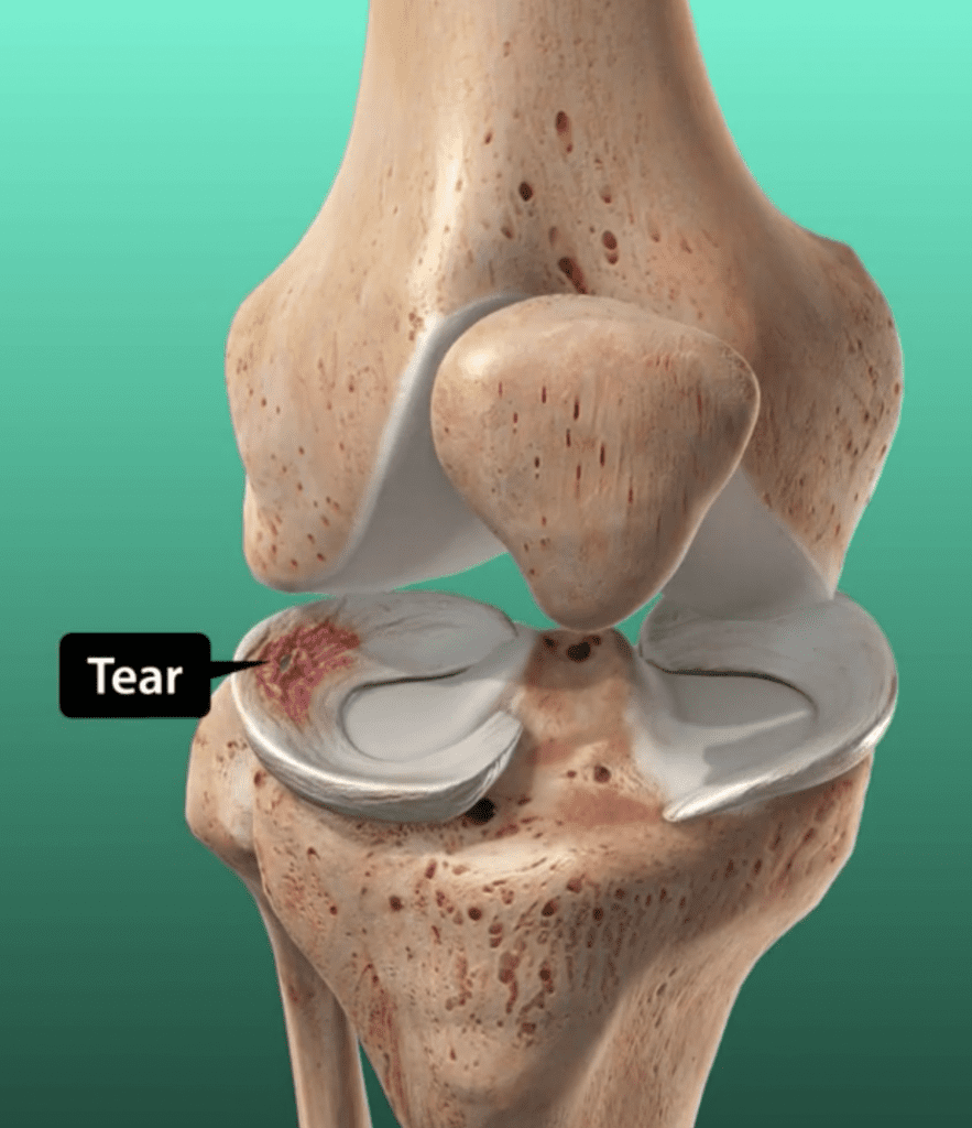
The knee joint, a marvel of biomechanical engineering, relies on several crucial components for its stability and smooth function. Among the most vital are the menisci – two C-shaped pieces of cartilage that act as cushions between your femur (thighbone) and tibia (shinbone). These resilient structures play an important role in absorbing force, distributing weight, and assisting in the nourishment of the knee joint. When healthy, they allow for seamless movement, but an injury can cause altered biomechanics of the knee joint and significant pain. Meniscus tears are, in fact, the most common injury of the knee [1]. While many people refer to them simply as "torn cartilage in the knee," they specifically involve these vital shock absorbers. Tears can occur in various ways: in younger patients, they often result from acute knee injuries, typically involving a sudden twist or rotation during physical activity. Athletes, particularly those involved in contact sports, are at a higher risk due to the dynamic forces exerted on their knees. However, significant trauma is not always necessary; a sudden twist or repeated squatting can tear the meniscus, making it a common injury for anyone at any age. In older individuals, meniscal tears may occur as part of a degenerative process, where the cartilage weakens over time, making it more susceptible to tearing even with minimal stress. Interestingly, medial meniscus tears are generally seen more frequently than tears of the lateral meniscus, with a ratio of approximately 2:1 [2]. Understanding the nature and prevalence of these injuries sets the stage for appreciating the importance of an accurate **meniscus injury test**.The Crucial Role of Diagnostic Tests in Meniscus Injury Assessment
When knee pain strikes, especially after a specific incident or with persistent discomfort, the immediate concern is often "What's wrong?" While symptoms like swelling, stiffness, and difficulty extending or bending the knee can point towards a meniscus injury, self-diagnosis is never sufficient. This is where specialized diagnostic tests come into play. These clinical examinations are the first line of defense in identifying a potential meniscus tear, allowing healthcare providers to gather crucial information about the injury before considering more advanced imaging. The primary goal of a **meniscus injury test** is to reproduce the patient's pain or symptoms by stressing the meniscus in specific ways. By carefully manipulating the knee and leg, a skilled examiner can often elicit signs that indicate a tear, such as a palpable click, pop, or localized pain. These tests are invaluable because they are non-invasive, relatively quick, and can provide immediate insights into the integrity of the menisci. They help healthcare providers narrow down the possibilities, guiding the diagnostic process efficiently and effectively. While these physical tests are highly indicative, they are often used in conjunction with a thorough patient history and, if necessary, confirmed by imaging studies like MRI.The McMurray Test: A Cornerstone of Meniscus Injury Diagnosis
Among the various orthopedic special tests used for physical diagnosis of knee lesions, the McMurray test stands out as the most common and widely recognized **meniscus injury test**. It's a classic examination that has been a staple in clinical practice for decades, providing valuable insights into the integrity of both the medial and lateral menisci.What is the McMurray Test?
The McMurray test is a series of knee and leg movements healthcare providers use to diagnose a torn meniscus. It is specifically designed to assess the integrity of the medial and lateral meniscus, testing for meniscal tears, which, as we know, are the most common injury to the knee. Over 1 million Americans tear their meniscus each year, making this test a frequently performed diagnostic procedure. It's often used along with the joint line tenderness test to identify meniscal injury, providing a more comprehensive clinical picture. The essence of the McMurray test lies in its ability to trap a torn piece of meniscus between the femur and tibia, thereby reproducing the patient's symptoms.How to Perform the McMurray Test
Performing the McMurray test requires precision and a good understanding of knee anatomy. This is how to perform it: To perform the McMurray test, the patient should be relaxed in a supine position (lying on their back) as the examiner firmly supports the knee. The examiner then flexes the knee fully, bringing the heel towards the buttocks. With one hand placed on the medial side of the knee (along the joint line), the examiner grasps the patient's foot with the other hand. The test proceeds with specific movements to target each meniscus:- To test the medial meniscus: The examiner externally rotates the leg (turns the foot outwards) while applying a valgus stress (pushing the knee inwards). While maintaining this position, the knee is slowly brought into extension (straightened) from full flexion.
- To test the lateral meniscus: The examiner internally rotates the leg (turns the foot inwards) while applying a varus stress (pushing the knee outwards). While maintaining this position, the knee is slowly brought into extension from full flexion.
Interpreting McMurray Test Results
The interpretation of the McMurray test is crucial for an accurate diagnosis. A positive test is indicated by pain, clicking, or popping within the knee joint as the knee is brought from flexion to extension. Specifically, a palpable pop or click combined with pain is considered a positive test and can correlate with a medial meniscus tear if elicited during external rotation and extension, or a lateral meniscus tear if elicited during internal rotation and extension. The test is considered positive for a meniscus tear if the patient experiences medial or lateral joint line discomfort or a sense of locking or catching in the knee. This "locking" or "catching" sensation often signifies a displaced fragment of the torn meniscus getting caught within the joint. The location of the pain and the specific movement that elicits the positive sign help the healthcare provider determine which meniscus (medial or lateral) is likely injured. The reliability of this test, when performed correctly, makes it an indispensable part of the clinical assessment for a suspected meniscus injury.Clinical Significance and Complementary Tests
The McMurray test of the knee is used to evaluate the injury of the lateral and medial meniscus of the knee joint. Its significance lies in its ability to provide immediate, actionable information during a physical examination. While it is a highly regarded **meniscus injury test**, it is rarely used in isolation. As mentioned, it's commonly paired with the joint line tenderness test, where the examiner gently palpates along the joint line to identify areas of localized pain, which can also indicate a meniscus tear. Orthopedic exams and special tests for physical therapy are designed to be comprehensive, and the McMurray test is just one piece of the diagnostic puzzle. It helps guide the next steps in patient care, whether that involves conservative management or further imaging. The ability of this test to reproduce symptoms that are characteristic of a torn meniscus makes it an invaluable tool for clinicians in their quest to accurately diagnose and manage knee injuries.The Thessaly Test: Another Valuable Tool for Meniscus Injury Detection
While the McMurray test is widely known, the Thessaly test is another important **meniscus injury test** that healthcare providers may use, especially when a patient's symptoms are more subtle or if the McMurray test is inconclusive. This dynamic test attempts to replicate the rotational forces that often lead to meniscus tears, making it particularly effective in certain scenarios.What is the Thessaly Test?
The Thessaly test is a series of knee and leg movements healthcare providers use to diagnose a torn meniscus. Unlike the McMurray test, which is performed with the patient supine, the Thessaly test is performed with the patient standing, making it a weight-bearing maneuver. The patient stands on the affected leg with the knee slightly flexed (typically at 5 degrees and then at 20 degrees). While maintaining this slight flexion, the patient rotates their body and the knee internally and externally three times. This test must be performed keeping in view the balance and pain of the patient. Because it's a weight-bearing test, it can be more provocative for certain types of meniscus tears, especially those that cause symptoms during activities like twisting or pivoting. A positive Thessaly test is indicated by medial or lateral joint line pain, or a sense of locking or catching in the knee during the rotational movements. The Thessaly test can be a valuable complement to other clinical examinations, offering an alternative way to stress the meniscus and elicit symptoms, thereby enhancing the accuracy of a **meniscus injury test** battery.Beyond Clinical Examination: Advanced Imaging for Confirmation
While physical examination tests like the McMurray and Thessaly tests are highly valuable initial diagnostic tools for a suspected **meniscus injury test**, they are not always definitive. Clinical tests provide strong indications, but for a confirmed diagnosis and to rule out other associated injuries, advanced imaging is often necessary. This is where Magnetic Resonance Imaging (MRI) plays a crucial role. MRI is a highly sensitive imaging modality that provides detailed images of soft tissues, including the menisci, ligaments, and cartilage within the knee joint. Reliability of test and other clinical tests like MRI is used to confirm the diagnosis of meniscal tear. MRI is sensitive, and it identifies associated injury. It can clearly show the location, type, and extent of a meniscus tear, as well as detect any other damage to surrounding structures like ligaments or articular cartilage. This comprehensive view is invaluable for surgical planning, if needed, and for determining the most appropriate course of treatment. While physical tests help guide the initial assessment, an MRI offers the definitive visual evidence that confirms the presence of a tear and provides a complete picture of the knee's internal condition, ensuring the most accurate diagnosis and treatment plan for a **meniscus injury test**.What to Do If You Suspect a Meniscus Tear
Discovering you might have a torn meniscus can be concerning, but knowing the right steps to take can make a significant difference in your recovery journey. Here’s what to do if your meniscus tears, or if you suspect it has: First and foremost, it's crucial to seek professional medical evaluation. While this article provides extensive information on the **meniscus injury test**, self-diagnosis is never recommended. A healthcare provider, such as an orthopedic specialist or sports medicine physician, is best equipped to accurately diagnose your condition. They will conduct a thorough physical examination, including the McMurray test and potentially the Thessaly test, and take a detailed medical history. In the immediate aftermath of a suspected injury, you can follow the R.I.C.E. protocol to manage symptoms:- Rest: Avoid activities that aggravate your knee pain. Give your knee a break from weight-bearing and twisting movements.
- Ice: Apply ice packs to the affected area for 15-20 minutes several times a day to reduce swelling and pain.
- Compression: Use an elastic bandage or compression sleeve to help minimize swelling.
- Elevation: Elevate your knee above the level of your heart whenever possible to reduce fluid accumulation.
Preventing Meniscus Injuries: Practical Tips
While not all meniscus tears can be prevented, especially those resulting from degenerative processes or unavoidable acute trauma, there are several practical steps you can take to significantly reduce your risk. Understanding these preventive measures can empower you to protect your knees and maintain their long-term health.- Warm-Up Properly: Before any physical activity, especially sports or exercises involving the knees, perform a thorough warm-up. This increases blood flow to your muscles and prepares your joints for activity, making them more pliable and less prone to injury.
- Strengthen Your Legs: Strong thigh and calf muscles provide better support and stability for your knee joint. Incorporate exercises like squats, lunges, and hamstring curls into your routine. Focus on balanced strength between your quadriceps and hamstrings.
- Improve Flexibility: Good flexibility in your hamstrings, quadriceps, and calf muscles can help prevent excessive stress on your knees during movement. Regular stretching can improve your range of motion.
- Use Proper Technique: Whether you're lifting weights, playing sports, or performing daily tasks, pay attention to your body mechanics. Learn and use correct form for exercises and movements. For instance, when squatting, ensure your knees don't go past your toes.
- Avoid Sudden Twists and Pivots: Many meniscus tears occur from forceful twisting or rotating movements, especially when the foot is planted. Be mindful of these movements, particularly during sports. If you need to change direction, try to pivot on the balls of your feet rather than twisting through your knee.
- Wear Appropriate Footwear: Ensure your shoes provide adequate support and cushioning for your activities. Worn-out or ill-fitting shoes can alter your gait and place undue stress on your knees.
- Listen to Your Body: Don't push through pain. If you feel discomfort in your knee, stop the activity and rest. Ignoring pain can lead to more severe injuries.
- Maintain a Healthy Weight: Excess body weight places additional stress on your knee joints, increasing the risk of both acute injuries and degenerative changes to the meniscus.
Conclusion
Navigating the complexities of knee pain can be daunting, but understanding the diagnostic journey, particularly the role of a thorough **meniscus injury test**, is a crucial first step toward recovery. We've explored how the menisci serve as vital shock absorbers in the knee and how their tears are incredibly common, affecting millions and often stemming from sudden twists or degenerative processes. The McMurray test, with its precise movements and characteristic positive signs like pops, clicks, and pain, remains the most common orthopedic special test, offering immediate insights into potential tears. Complementing this, the Thessaly test provides another valuable diagnostic avenue, especially in weight-bearing scenarios. While these clinical examinations are foundational, advanced imaging like MRI offers definitive confirmation and a comprehensive view of any associated injuries. Ultimately, suspecting a meniscus tear necessitates prompt medical attention for an accurate diagnosis and tailored treatment plan. We hope this comprehensive guide has demystified the process of diagnosing a meniscus injury, empowering you with knowledge and confidence. If you've found this information helpful, consider sharing it with others who might benefit. Do you have experiences with a meniscus injury or these diagnostic tests? Share your thoughts and questions in the comments below – your insights could help others on their journey to knee health!- Xxxxxx Is Equal To 2 X 5
- Sone 793
- Remote Access Raspberry Pi From Internet
- Soviet Seduction Jackerman
- Jayshree Gaikwad Movies
Anatomy Of The Knee Diagram Torn Meniscus

Torn Meniscus Trimming – UnderstandOrtho™
Anatomy Of Medial Meniscus
